Cutting edge 3D skull model aids jaw lock surgery for cancer survivor
Doctors in the city generated a cutting-edge three-dimensional model of a skull of a 31-year-old cancer survivor, who had been on a liquid diet for more than a year and weighed only 40 kgs.
She struggled with a complete jaw lock, impeding her ability to open her mouth.
Sets of MRI and CT scans helped to create the replica for thorough examination before proceeding with temporomandibular joint replacement surgery.
At 31, Saba Shaikh encountered considerable challenges in opening her mouth and consuming food due to stiffness in her temporomandibular joint (TMJ)– where the jaw connects to the skull, facilitating jaw movement for chewing and speaking.
Relying on a liquid diet for over a year, she resorted to using a flat spoon for eating. This gradual decline in her ability to eat normally not only impacted her physical health but also took a toll on her mental well-being, hindering her speech clarity. “We visited several hospitals and she underwent an open joint surgery of TMJ to address the issue. Unfortunately, this procedure proved unsuccessful and resulted in Ankylosis (abnormal stiffening) of the TMJ,” said her husband Azharuddin Shaikh.
The Navi Mumbai resident was diagnosed with nasopharyngeal cancer, a type of head and neck cancer that originates in the nasopharynx (the area behind the nose) four years ago. With radiation and chemotherapy, she successfully recovered.
Afterwards, she began experiencing TMJ pain and struggled to open her mouth. Over time, her ability to open her mouth decreased, resulting in complete lockjaw. This led to significant weight loss and dental issues, including tooth decay and gum swelling, due to compromised oral hygiene.
“Her mouth opening had further decreased, to less than the width of one finger, with only the right joint functioning and the left joint completely ankylosed (becoming stiff),” said Azharuddin.
Later, she approached Dr Tofiq Bohra, consultant, oral and maxillofacial surgery department at SL Raheja Hospital. Dr Bohra conducted an examination and subsequently ordered CT and MRI scans of both sides of her jaw joint. The results indicated stiffness and erosion on the left side and wear on the right side.
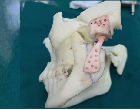
After a thorough analysis of the patient’s CT scan and MRI findings, a 3D replica of her skull was developed. The medical team recommended TMJ replacement surgery, to which Saba agreed.
The design of the implant was then created using a 3D printer. On March 26, the TMJ prosthesis was implanted. To provide additional protection to the prosthesis, it was covered with a layer of the patient’s abdominal fat.
“TMJ replacement surgery offers an advanced solution, surpassing alternative methods by implanting a new prosthetic joint in the facial area, reducing the risk of Ankylosis recurrence,” said Dr Bohra.
“One challenge faced was inserting an anaesthesia breathing tube due to restricted mouth opening, but this was successfully addressed through nasal endoscopy with an anaesthetist’s assistance. Furthermore, the surgery was complicated by the presence of fibrosis resulting from prior joint replacement procedures,” added Dr Bohra.
Within 48 hours of the surgery, her mouth opened up to two fingers wide. Currently, Sana is on physiotherapy and on a soft diet. “Finally, after months, my wife is able to eat her favourite foods,” said Azharuddin.
© The Indian Express Pvt Ltd